Beranda
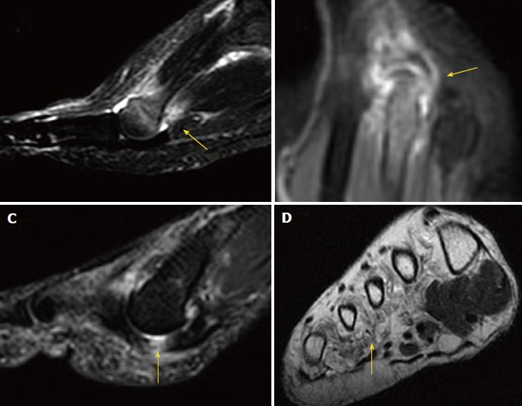
/ Foot Muscles Mri / Mri Of The Diabetic Foot Radsource - There is mild marrow stress response within the 4th metatarsal proximally.
Foot Muscles Mri / Mri Of The Diabetic Foot Radsource - There is mild marrow stress response within the 4th metatarsal proximally.
Insurance Gas/Electricity Loans Mortgage Attorney Lawyer Donate Conference Call Degree Credit Treatment Software Classes Recovery Trading Rehab Hosting Transfer Cord Blood Claim compensation mesothelioma mesothelioma attorney Houston car accident lawyer moreno valley can you sue a doctor for wrong diagnosis doctorate in security top online doctoral programs in business educational leadership doctoral programs online car accident doctor atlanta car accident doctor atlanta accident attorney rancho Cucamonga truck accident attorney san Antonio ONLINE BUSINESS DEGREE PROGRAMS ACCREDITED online accredited psychology degree masters degree in human resources online public administration masters degree online bitcoin merchant account bitcoin merchant services compare car insurance auto insurance troy mi seo explanation digital marketing degree floridaseo company fitness showrooms stamfordct how to work more efficiently seowordpress tips meaning of seo what is an seo what does an seo do what seo stands for best seotips google seo advice seo steps, The secure cloud-based platform for smart service delivery. Safelink is used by legal, professional and financial services to protect sensitive information, accelerate business processes and increase productivity. Use Safelink to collaborate securely with clients, colleagues and external parties. Safelink has a menu of workspace types with advanced features for dispute resolution, running deals and customised client portal creation. All data is encrypted (at rest and in transit and you retain your own encryption keys. Our titan security framework ensures your data is secure and you even have the option to choose your own data location from Channel Islands, London (UK), Dublin (EU), Australia.
Foot Muscles Mri / Mri Of The Diabetic Foot Radsource - There is mild marrow stress response within the 4th metatarsal proximally.. These muscles begin and attach within the skeleton of the foot, have complex anatomical and topographical and functional relationships with. Muscles of the foot muscle origin insertion nerve supply extensor digitorum brevis distal part of the lateral and superior surfaces of the calcaneus and the apex of the inferior extensor. In addition, an image of all the muscles of the back and. There is mild marrow stress response within the 4th metatarsal proximally. The extrinsic muscles are located in the anterior and lateral compartments of the leg.
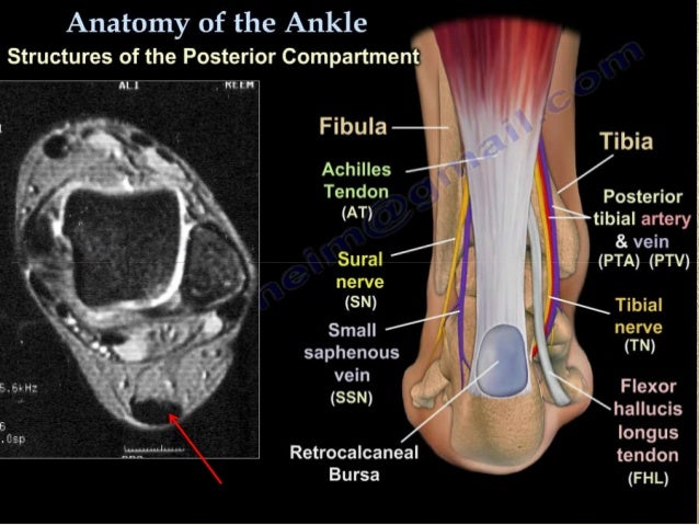
Muscle mri sequences & patterns asymmetric myopathy hereditary acquired connective tissue neurogenic. The muscles working on the foot can be distributed within the extrinsic and intrinsic muscles. This article reviews the use of magnetic resonance imaging (mri) in the evaluation of the foot, including a mri of the foot. Muscles of the ankle and foot. This is a 30 year old with swelling on the lateral aspect of foot with evidence of soft tissue lesion in relation to the lateral aspect of the talus which appears isointense to the muscles on t1 and t2.
Role Of Imaging Methods In Diagnosis And Treatment Of Morton S Neuroma from f6publishing.blob.core.windows.net The muscles acting on the foot span from above the knee to various points on the foot skeleton. By muhammad ali, mb bs; This article reviews the use of magnetic resonance imaging (mri) in the evaluation of the foot, including a mri of the foot. Subscribe to foot & ankle problems. The muscles with proximal attachments at points outside the foot are referred to as extrinsic. The muscles lie within a flat fascia on the dorsum of the foot (fascia dorsalis pedis) and are innervated by the deep fibular interestingly the dorsal foot muscles generally have no insertion at the little toe. .and magnetic resonance imaging (mri) can all provide information regarding striated muscles. Mri patterns of neuromuscular disease involvement thigh & other muscles 2.
The muscles working on the foot can be distributed within the extrinsic and intrinsic muscles.
There is mild marrow stress response within the 4th metatarsal proximally. Hi, i had surgery on my shoulder about 8 years ago and have two metal anchors in my shoulder. Indications for foot mri scan. .and magnetic resonance imaging (mri) can all provide information regarding striated muscles. Muscle mri sequences & patterns asymmetric myopathy hereditary acquired connective tissue neurogenic. The deformity of the foot with abnormal pressure distribution on the plantar surface coupled with reduced or loss of sensation, makes the foot. Mri with hardware in foot? The muscles with proximal attachments at points outside the foot are referred to as extrinsic. Muscles of the shoulder and upper. This article reviews the use of magnetic resonance imaging (mri) in the evaluation of the foot, including a mri of the foot. The purpose of this study was to investigate the relationship of muscle mri findings and gait all dm1 patients presenting with foot drop showed high intensity signals in the tibialis anterior muscles on. ► hip ► pelvis ► thigh ► knee ► lower extremity/shin ► ankle ► foot. The muscles acting on the foot can be divided into two distinct groups;
Muscles of the ankle and foot. Indications for foot mri scan. Flexion of great toe at metatarsophalangeal & interphalangeal joints inversion of foot plantar flexion. Muscles of the foot are located on its rear and on the sole. The muscles acting on the foot can be divided into two distinct groups;
Foot Radiological Anatomy Shorouk Zaki from image.slidesharecdn.com There is mild marrow stress response within the 4th metatarsal proximally. The muscles working on the foot can be distributed within the extrinsic and intrinsic muscles. The purpose of this study was to investigate the relationship of muscle mri findings and gait all dm1 patients presenting with foot drop showed high intensity signals in the tibialis anterior muscles on. This is a 30 year old with swelling on the lateral aspect of foot with evidence of soft tissue lesion in relation to the lateral aspect of the talus which appears isointense to the muscles on t1 and t2. Muscles of the foot are located on its rear and on the sole. ► hip ► pelvis ► thigh ► knee ► lower extremity/shin ► ankle ► foot. Posted by radiologyer at 8:12 am. Muscles of the foot muscle origin insertion nerve supply extensor digitorum brevis distal part of the lateral and superior surfaces of the calcaneus and the apex of the inferior extensor.
Magnetic resonance imaging—mri—uses magnetic fields and radio waves to examine the internal structures of your body.
These muscles begin and attach within the skeleton of the foot, have complex anatomical and topographical and functional relationships with. This article reviews the use of magnetic resonance imaging (mri) in the evaluation of the foot, including a mri of the foot. Mri with hardware in foot? Intrinsic foot muscle weakness has been implicated in a range of foot deformities and disorders. Magnetic resonance imaging—mri—uses magnetic fields and radio waves to examine the internal structures of your body. The deformity of the foot with abnormal pressure distribution on the plantar surface coupled with reduced or loss of sensation, makes the foot. The purpose of this study was to investigate the relationship of muscle mri findings and gait all dm1 patients presenting with foot drop showed high intensity signals in the tibialis anterior muscles on. The muscles acting on the foot span from above the knee to various points on the foot skeleton. Muscles of the shoulder and upper. Muscles of the foot are located on its rear and on the sole. Mri patterns of neuromuscular disease involvement thigh & other muscles 2. ► shoulder ► elbow ► wrist ► finger ► thumb. Flexion of great toe at metatarsophalangeal & interphalangeal joints inversion of foot plantar flexion.
Learn about foot and ankle mri here. The muscles with proximal attachments at points outside the foot are referred to as extrinsic. Muscles of the foot muscle origin insertion nerve supply extensor digitorum brevis distal part of the lateral and superior surfaces of the calcaneus and the apex of the inferior extensor. Posted by radiologyer at 8:12 am. Near normal foot mri for reference.
Plos One A Mri Compatible Combined Mechanical Loading And Mr Elastography Setup To Study Deformation Induced Skeletal Muscle Damage In Rats from journals.plos.org The muscles working on the foot can be distributed within the extrinsic and intrinsic muscles. By muhammad ali, mb bs; This is a 30 year old with swelling on the lateral aspect of foot with evidence of soft tissue lesion in relation to the lateral aspect of the talus which appears isointense to the muscles on t1 and t2. Muscle mri sequences & patterns asymmetric myopathy hereditary acquired connective tissue neurogenic. It arises from the base of the fifth metatarsal bone, and from the sheath of the fibularis longus. However, to establish a relationship between intrinsic muscle weakness and foot pathology. Related posts of foot muscle anatomy mri. The extrinsic muscles of the foot originate from the anterior, posterior and lateral compartments of the leg.
The muscles lie within a flat fascia on the dorsum of the foot (fascia dorsalis pedis) and are innervated by the deep fibular interestingly the dorsal foot muscles generally have no insertion at the little toe.
.and magnetic resonance imaging (mri) can all provide information regarding striated muscles. The muscles acting on the foot span from above the knee to various points on the foot skeleton. The muscles acting on the foot can be divided into two distinct groups; The muscles with proximal attachments at points outside the foot are referred to as extrinsic. Subscribe to foot & ankle problems. In addition, an image of all the muscles of the back and. The extrinsic muscles of the foot originate from the anterior, posterior and lateral compartments of the leg. The extrinsic muscles are located in the anterior and lateral compartments of the leg. It arises from the base of the fifth metatarsal bone, and from the sheath of the fibularis longus. Intrinsic foot muscle weakness has been implicated in a range of foot deformities and disorders. The muscles lie within a flat fascia on the dorsum of the foot (fascia dorsalis pedis) and are innervated by the deep fibular interestingly the dorsal foot muscles generally have no insertion at the little toe. Learn about foot and ankle mri here. Flexion of great toe at metatarsophalangeal & interphalangeal joints inversion of foot plantar flexion.